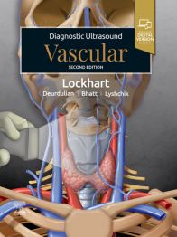
Diagnostic Ultrasound: Vascular - E-book, 2nd Edition
Develop a solid understanding of ultrasound and evolving vascular ultrasound practices with this practical, point-of-care reference in the popular Diagnostic Ultrasound series. Written by leading experts in the field, the second edition of Diagnostic Ultrasound: Vascular offers detailed, clinically oriented coverage of anatomy, techniques, and diagnoses in this complex area. Featuring more than 1,750 images and full-color illustrations throughout, this edition showcases vascular ultrasound techniques across 4 different types of ultrasound, including details regarding imaging artifacts. Diagnostic pearls and pitfalls accompany the detailed sonographic descriptions of vascular disease and anomalies regularly encountered in the head and neck, chest and abdomen (including transplants), and extremities.
Key Features
- Provides a wide range of anatomic detail, technical factors, and diagnostic criteria to guide accurate application of ultrasound throughout the body
- Covers new and evolving techniques such as the increasing use of microbubble imaging to enhance image resolution, distinguish vessels more clearly, and minimize noise and background signals
- Details the latest information across several ACR RADS criteria, and contains extensive new material from the LI-RADS, GB-RADS, and transplant criteria, which now include Doppler ultrasound with its noninvasive methodology rated highly for appropriate use
- Reflects an increased use of Doppler extremity evaluations due to ongoing COVID-19 diagnoses and a higher incidence of venous thrombosis
- Contains updated ACR Appropriateness Criteria regarding the new “highly appropriate” ratings, as well as new Intersocietal Accreditation Commission (IAC) recommendations in numerous diagnosis chapters
- Contains a gallery of typical and atypical ultrasound appearances covering a wide spectrum of disease, correlated with CT and MR imaging where appropriate, and detailed artistic renderings
- Features image-rich chapters on vascular ultrasound techniques, covering grayscale, color, power, and spectral (pulsed) Doppler imaging, as well as imaging artifacts
- Contains time-saving reference features such as succinct and bulleted text, a variety of test data tables, a Key Facts section that begins in each chapter, annotated images, and an extensive index
- An ideal reference for radiologists, sonographers, vascular surgeons, and those who are training in these fields
Author Information
| ISBN Number | 9780443283673 |
|---|---|
| Main Author | By Mark E. Lockhart, MD, MPH |
| Copyright Year | 2025 |
| Edition Number | 2 |
| Format | eBook |
| Illustrations | 1823 full-color images and illustrations |
| Imprint | Elsevier |
| Page Count | 630 |
| Publication Date | 13 Sep 2024 |
| Stock Status | IN STOCK |


eBooks : Built for busy schedules & tailored for your goals.


Affordable knowledge, built for you
Get the resources you need-often at a lower cost than print. Quality content designed to support your goals, without stretching your budget.


Seamless access wherever you are


Always in sync


Tools that make learning stick
Your eBook is ready whenever you are!
1. Check your email for your access code.


2. Sign into or create your VitalSource account and redeem your code.


3. Open your eBook - ready whenever you are!
FAQ
The access code for your new eBook will be sent in your order confirmation email. Your code can also be accessed in your My Account section on the Elsevier webshop. If you do not receive your code within a few minutes, please check your spam folder.
Step-by-step guidance on how to download Bookshelf and also redeem your code can be found here.
The access code for your new eBook does not expire. However, we always suggest redeeming immediately after purchase to start experiencing the benefits of and insights from your purchase. Important to note - the code provided is a single use code and only valid for the edition you purchase. It does not provide access to past nor future editions of the title.
You will have unlimited access to your eBook on the device to which it was downloaded.
Discover the various learning features that our eBooks offer on the Bookshelf® Reader! For example, you can highlight different text passages, create notes and flashcards, have the text read to you, etc. Particularly practical: You can also use your eBooks offline. More information on the learning functions can be found on the Vitalsource page.
Quality is our top priority. That's why we collaborate with the leading eBook reader provider VitalSource. VitalSource has its own eBook reader Bookshelf®, which you can easily download. This reader is very user-friendly and offers more features than other standard readers. For example, you can highlight different text passages, create notes and flashcards, have the text read to you, etc. Particularly practical: You can also use your eBooks offline. More information can be found on the Vitalsource page.
Elsevier offers its eBooks in ePub format, as we believe this format is best suited to display our content ideally on as many devices as possible.
You cannot return eBooks. If you have any questions, feel free to contact us.
Any questions ?
Top Picks from Our Community
eBook
eBook
Shadi Khalil
Aug 2026
eBook
Edited by Albert J. Heuer
Aug 2026
eBook
eBook
Edited by Michael P. Steinmetz
Jul 2026
eBook
Brittany Hunter
Jul 2026
eBook
Edited by Roberta L. Hines
Jul 2026
eBook
eBook
Gregory J. Hughes
Jul 2026
eBook
eBook
Edited by Wesley S. Moore
Jul 2026
eBook
Kambiz Motamedi
Jul 2026
eBook
Shelly Rainforth Collins
Jul 2026
eBook
Edited by Robert H. Anderson
Jul 2026
eBook
eBook
Edited by Mathew Chung
Jul 2026
eBook
Edited by Joy E. Tomlinson
Jul 2026
eBook
Edited by Neil J. Friedman
Jul 2026
eBook
Edited by Seth E. Kaplan
Jul 2026
eBook
Edited by Katherine P. Liao
Jul 2026
eBook
Edited by Natalia Nagornaya
Jul 2026
eBook
Lynn B. Jorde
Jul 2026
eBook
Edited by Waseem Anani
Jul 2026
eBook
eBook
Steven A. Cole
Jul 2026
Part I: Technique
Section 1: Modalities
Color Doppler
Power Doppler
Spectral Doppler: General Waveform Concepts
Contrast-Enhanced Ultrasound: Basic Technique in Liver and Kidney
Volumetric Blood Flow
Microvascular Imaging
Section 2: Artifacts
Aliasing, Blooming, and Twinkling Artifact
Part II: Anatomy
Section 1: Head and Neck
Neck Overview
Cervical Carotid Arteries
Vertebral Arteries
Neck Veins
Section 2: Chest and Abdomen
Thoracic Outlet
Ribs and Intercostal Space
Liver
Spleen
Pancreas
Kidneys
Aorta and Inferior Vena Cava
Mesenteric Vessels
Iliac Arteries and Veins
Penile
Scrotum
Gynecologic Structures
Section 3: Extremities
Upper Limb
Arm Overview
Arm Vessels
Forearm Overview
Wrist Overview
Hand Overview
Wrist and Hand Vessels
Lower Limb
Groin Overview
Femoral Vessels and Nerves
Lower Leg Overview
Lower Leg Vessels
Foot Vessels
Part III: Diagnoses
Section 1: Head and Neck
Parotid Vascular Lesion
Venous Vascular Malformation
Jugular Vein Thrombosis
Carotid Artery Dissection in Neck
Carotid Stenosis/Occlusion
Carotid-Jugular Fistula
Vertebral Stenosis/Occlusion
Atypical Carotid Waveforms
Arteritis/Takayasu
IABP and Other Devices
Section 2: Chest and Abdomen
Aorta and Vena Cava
Aortic/Iliac Aneurysm
Aortoiliac Occlusive Disease
IVC Obstruction
Aortic Dissection
Aortic Stent Endoleak
Celiac Artery Stenosis
SMA Stenosis
Median Arcuate Ligament Syndrome
Liver
Portal Hypertension
Approach to Hepatic Sonography
Portosystemic Collaterals
Transjugular Intrahepatic Portosystemic Shunt (TIPS)
Portal Vein Occlusion
Budd-Chiari Syndrome
Portal Vein Gas
Hepatic Tumor Thrombus/Tumor-in-Vein
Spleen
Splenic Vascular Disorders
Splenic Artery Aneurysm
Splenic Vein Thrombosis
Kidney
Renal Artery Stenosis
Renal Vein Thrombosis
Renal Infarct
Perinephric Hematoma
Tumor Vascularity
Renal AVF/AVM
Renal Vein Nutcracker
High Resistive Index
Reproductive
Varicocele
Testicular Torsion/Infarct
Penile
Ovarian Torsion
Uterine AVM/PSA
Uterine Fibroid Embolization
Pelvic Congestion
Section 3: Transplants
Kidney
Approach to Sonography of Renal Allografts
Transplant Renal Artery Stenosis
Transplant Renal Artery Thrombosis
Transplant Renal Vein Thrombosis
Renal Transplant Arteriovenous Fistula
Renal Transplant Pseudoaneurysm
Renal Transplant Rejection
Delayed Renal Graft Function
Liver
Liver Transplant Hepatic Artery Stenosis/Thrombosis
Liver Transplant Portal Vein Stenosis/Thrombosis
Liver Transplant Hepatic Venous Stenosis/Thrombosis
Liver Transplant Biliary Stricture
Hepatic Artery Aneurysm/Pseudoaneurysm
Pancreas
Pancreas Transplant: Normal vs. Rejection
Section 4: Extremities
Arteries
Peripheral Arterial Occlusive Disease
Peripheral Arterial Pseudoaneurysm
Peripheral Arteriovenous Fistula
Peripheral Artery Bypass
Wrist Modified Allen Test
Hemodialysis Stenosis/Occlusion
Hemodialysis Failure to Mature
Hemodialysis Steal
Veins
Deep Vein Thrombosis
Varicose Veins/Incompetent Perforator
Iliac and IVC Venous Occlusion
Soft Tissues
Limb Hemangioma
Vascular Dilatation or Inflammation
Vascular Anomaly
Vascular Tumor/Leiomyosarcoma
Section 5: Interventions
Thrombin PSA Repair
Aneurysm Coiling