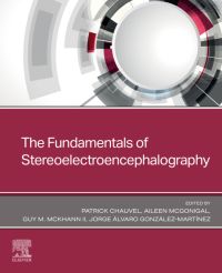
Gain confidence and expertise with stereoelectroencephalography (SEEG) as a presurgical method for epilepsy with this helpful resource. Edited and written by leading experts in the field, Stereoelectroencephalography teaches the scientific and medical bases of SEEG through its essential disciplines (anatomy, biophysics, electrophysiology, and cognitive and behavioral neuroscience) and their interrelations. It fully covers the basic and clinical aspects of the pharmaco-resistant epilepsies investigated with SEEG and their surgical indications.
Key Features
-
Describes the evolution in time of the presurgical methods leading to the current practice of SEEG.
-
Explains how to determine the anatomical basis of electrode implantation, its referential system, and how it prepares rational planning to tailored resection or ablation.
-
Examines the nature of the SEEG signal, how the depth electrodes capture it, and how the dynamics of multiple cortical sites recording can be understood.
-
Contains chapters on key topics such as the optimal SEEG electrode, intracerebral electrodes implantation technique, seizure onset: interictal, preictal/ictal patterns and the epileptogenic zone, how to define the extent of the EZ by applying signal processing methods, the role of SEEG in exploring lesional epilepsy cases, and much more.
-
Presents the logical chain linking SEEG to anatomically pre-planned surgery.
-
Includes discussions of in-depth illustrative cases.
Author Information
Edited by Patrick Chauvel, MD, Epilepsy Center, Neurological Institute, Cleveland Clinic, Cleveland, OH, United States; Jorge Álvaro Gonzalez-Martinez, MD, PhD, University of Pittsburgh Medical Center, Epilepsy & Movement Disorders Program, University of Pittsburgh Epilepsy Center, Cortical Systems Laboratory, UPMC Presbyterian, Pittsburgh, PA, United States; Aileen McGonigal, MBCHB, PhD, Department of Neurosciences, Mater Misericordiae Hospital, Brisbane, QLD, Australia; Mater Research Institute, Faculty of Medicine, University of Queensland, Brisbane, QLD, Australia; Queensland Brain Institute, University of Queensland, Brisbane, QLD, Australia and Guy M. McKhann II, MD, Department of Neurosurgery, Columbia University, New York, NY, United States


